Long Bone Diagram Red Marrow - The Skeletal System Biology For Majors Ii : There is a printable worksheet available for download here so you can take the quiz with pen and paper.
Long Bone Diagram Red Marrow - The Skeletal System Biology For Majors Ii : There is a printable worksheet available for download here so you can take the quiz with pen and paper.. Red marrow and yellow marrow. All red blood cells and platelets in adults are formed within red bone marrow, as well as 60% to 70% of white blood cells. The red color can be attributed to the hemoglobin. At birth, the whole skeleton is filled with red bone marrow. A typical long bone shows the gross anatomical characteristics of bone.
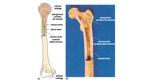
It can be found in the flat and long bones such as hip bones, vertebrae, ribs, shoulder blades, and skull. This type of bone marrow is found in your long bones and is usually surrounded. Marrow produces all types of blood cells and is where stem cells are found. The vertebrae and the long bones are bones with large marrow cavities,. A typical long bone showing gross anatomical features.
Epiphysis Wikipedia from upload.wikimedia.org There are actually two types of bone marrow: Red marrow is highly vascular, contains little fat and may be a source of blood cells of various types. This diagram depicts final long bone diagram. 4.8).compared to malignant bone marrow lesions, foci of red marrow have. The outer shell of the long bone is made of cortical bone also known as compact bone. Bone marrow is the soft, highly vascular and flexible connective tissue within bone cavities which serve as the primary site of new blood cell production or hematopoiesis. Found in the ends of long bones; A typical long bone shows the gross anatomical characteristics of bone.
There are actually two types of bone marrow:
It can be found in the flat and long bones such as hip bones, vertebrae, ribs, shoulder blades, and skull. Bone marrow is either red or yellow, depending upon the preponderance of hematopoietic (red) or fatty (yellow) tissue. The thigh bone (femur) is a long bone. This diagram depicts final long bone diagram. The red color can be attributed to the hemoglobin. It is essential for the body to function correctly. Hematopoietic is the formation and development of blood cells in the bone marrow. A typical long bone showing gross anatomical features. The structure of a long bone allows for the best visualization of all of the parts of a bone (figure 1). Red marrow fills the spaces in the spongy bone. This is an online quiz called long bone diagram. There are actually two types of bone marrow: Red bone marrow is also known as medulla osium rubra.
The red color can be attributed to the hemoglobin. Red bone marrow produces blood cells while yellow bone marrow stores fat. A typical long bone showing gross anatomical features. The structure of a long bone allows for the best visualization of all of the parts of a bone (figure 1). Bone marrow is either red or yellow, depending upon the preponderance of hematopoietic (red) or fatty (yellow) tissue.
1 716 Best Human Bone Marrow Images Stock Photos Vectors Adobe Stock from t3.ftcdn.net 4.8).compared to malignant bone marrow lesions, foci of red marrow have. A diagram of the anatomy of a bone, showing the bone marrow. Red bone marrow has stem cells that grow into red blood cells, white blood cells, and platelets. At birth, all bone marrow is red. Red marrow fills the spaces in the spongy bone. Bone marrow is either red or yellow, depending upon the preponderance of hematopoietic (red) or fatty (yellow) tissue. Spinal metastases initially involve the vertebral body. The main difference between red and yellow bone marrow is the occurrence and function of the each type of bone marrow in the body.
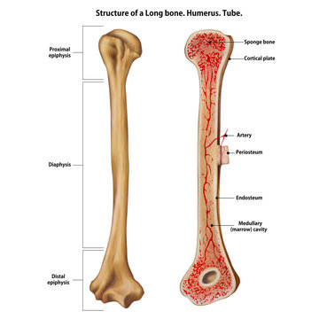
The diaphysis and the epiphysis.
The wider section at each end of the bone is called the epiphysis (plural = epiphyses), which is filled with spongy bone. A component of the lymphatic system, bone marrow functions primarily to produce blood cells and to store fat.bone marrow is highly vascular, meaning that it is richly supplied with a large number of blood vessels.there are two categories of bone marrow tissue: This diagram depicts final long bone diagram. In adults, all red marrow is found only in the proximal ends of the long bones of the limbs like the femur (as shown in the illustration) and in the breastbone, spine, ribs, shoulder blades, pelvis, and skull. The main difference between red and yellow bone marrow is the occurrence and function of the each type of bone marrow in the body. This is one of the two types of osseous tissue responsible for bone formation, and it is the softer and less rigid of the two. A typical long bone shows the gross anatomical characteristics of bone. In babies, all bone marrow is red. Bone marrow is the soft, highly vascular and flexible connective tissue within bone cavities which serve as the primary site of new blood cell production or hematopoiesis. There is a printable worksheet available for download here so you can take the quiz with pen and paper. In humans the red bone marrow forms all of the blood cells with the exception of the lymphocytes, which are produced in the marrow and reach their mature form in the lymphoid organs. The entire process is completed by the age of 25 years. Bone marrow is either red or yellow, depending upon the preponderance of hematopoietic (red) or fatty (yellow) tissue.
Red marrow fills the spaces in the spongy bone. 4.8).compared to malignant bone marrow lesions, foci of red marrow have. A diagram of the anatomy of a bone, showing the medullary cavity. At birth, the whole skeleton is filled with red bone marrow. There are actually two types of bone marrow:
Week 3 Tissue Structure And Function 2 3 Structure And Strength Functions Of Bone And Cartilage Openlearn Open University Oufl 008 from www.open.edu Bone marrow is the soft, highly vascular and flexible connective tissue within bone cavities which serve as the primary site of new blood cell production or hematopoiesis. A diagram of the anatomy of a bone, showing the medullary cavity. Bone marrow is the soft, flexible connective tissue within bone cavities. Half of it is converted to yellow marrow by age seven. The wider section at each end of the bone is called the epiphysis (plural = epiphyses), which is filled with spongy bone. It carries oxygen to the organs and helps prevent infections. A typical long bone showing gross anatomical features. There is a printable worksheet available for download here so you can take the quiz with pen and paper.
A long bone has two parts:
Red marrow fills the spaces in the spongy bone. The spine is the most common site for bone metastases because of the abundance of red marrow. 6 mesenchymal is embryonic tissue from which the connective tissue, blood vessels and lymphatic vessels are formed. The thigh bone (femur) is a long bone. 4.8).compared to malignant bone marrow lesions, foci of red marrow have. Red bone marrow helps produce blood cells; In an adult, hypointense marrow foci are a common finding, and they are often due to rests of hematopoietic marrow. Red marrow is composed of: Marrow produces 200 billion new blood cells daily. Spinal metastases initially involve the vertebral body. Physiological conversion of red into yellow bone marrow, lasting for the whole childhood until adulthood and has a constant, predictable pattern in the whole skeleton as well as in individual bones. Red marrow fills the spaces in the spongy bone. The outer shell of the long bone is made of cortical bone also known as compact bone.


Posting Komentar
0 Komentar